
Appliance for Immune- difusion test in agar wells
Visual control of interaction between antibody and anti-gene is known as "Immune- diffusion" reaction. It is generally used in research and scientific institutions. It is also indicated as DID (double immune diffusion), or Ouchterlony Double Diffusion.
Also, in experimental work or in cases of research of vaccinas to new virus, this kind of equipment is used with agar-fixation galss table. Special shape of lines after precipitation reaction render visual information about antibody and anti-gene interaction, and hence, effectiveness of vaccine.
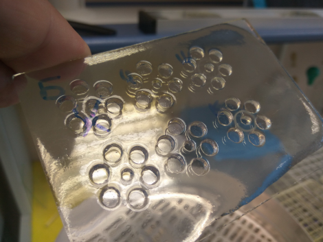
After hot agar is evenly spread in thin (2-3 mm) film, the researcher makes 7 perforation (wells) to be filled with serum (central well), and with buffer including varity of anti-dots - around in other six wells.
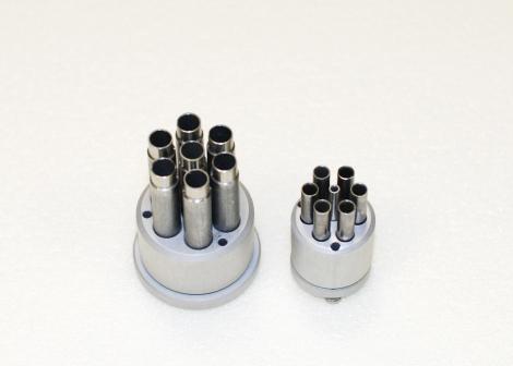
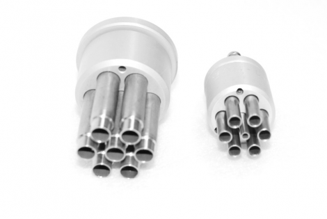
Two types (sizes) are produced, with 7.0 mm holes and 7 mm center well; other type - specially for brucellosis tests (according to standard method), having six 5 mm wells around 3 mm cental well.
After contact piercing of 7 wells in agar, he central part (circles) have to be removed by aspitation, or special vacuum pump. For inerpretation of the results, diffuse light source is needed.
More detailed description of method can be found at this video: https://www.youtube.com/watch?v=NsxdtxM1-qI
(Link is published with the kind permission of Frank Lectures).
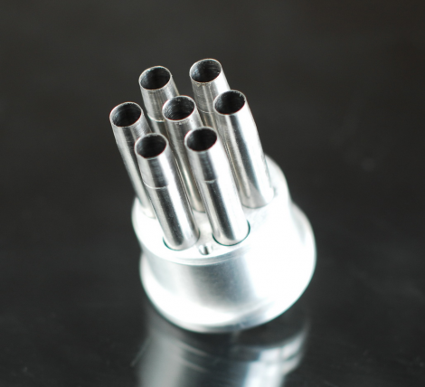
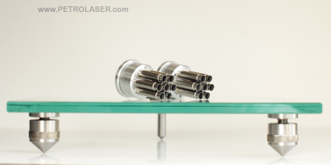
Perforation appliance is made of stainless steel and calibrated for precise cut- off the wells in Petri dish or glass plates. All- corrosion - free metal design.
Seven hollow tubes, 7 mm diam. each, with sharp edge. Mounted in equal distances in the metal holder.
Immune research, agar wells diffusion test: a diagnostic test using serum (the fluid, non-cellular part of blood) that detects antibody produced in response to infection. Serum is placed in a well in the agar and a MAP antigen preparation is placed in a nearby well. These two test components passively diffuse out of the well into the agar. If the serum sample contains antibodies to antigens of MAP they bind, forming an interlaced antigen-antibody complex that precipitates in the agar. The precipitate is visible to the unaided eye as a thin white line. This same technology is used for diagnosis of other diseases. The test is best interpreted by using a known positive control serum sample (provided in commercial kits) in the assay for comparison. For correct carrying out of this test, the small wells in agar should be in equal distances, and have equal sizes.
Can withstand hot sterilization (> 200C).
Details for usage can be found in this video: https://www.youtube.com/watch?v=oYnXeAPieN0
Reference to the video is published with kind permission of Food and Agriculture Organization of the United Nations.